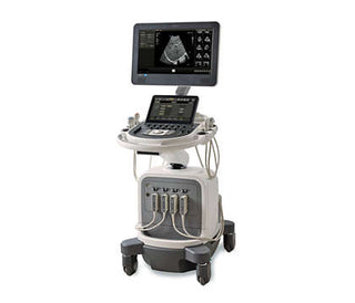
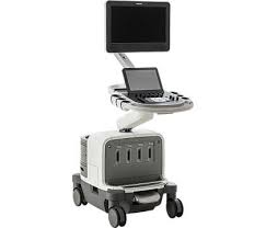
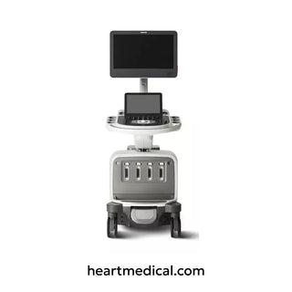
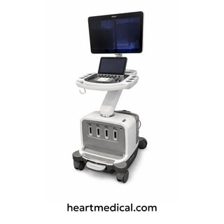
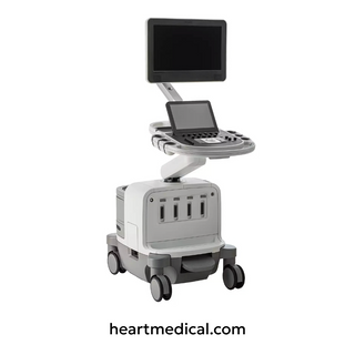
The Philips Epiq 5/7 is a premium ultrasound system designed for advanced diagnostic imaging. Introduced in the 2010s, it combines high-resolution imaging with user-friendly software, making it a favorite in hospitals and clinics.
Key Features
- PureWave Crystal Technology: Delivers clearer images, even in patients with challenging body types.
- Anatomical Intelligence: Automatically optimizes settings for specific organs.
- Versatility: Used for cardiac, abdominal, obstetric, vascular, and interventional procedures.
- Why it matters: The Epiq 5/7 helps doctors diagnose conditions faster and more accurately, improving patient outcomes.
List of All EPIQ 5/7 Compatible Probes

| Probe Model | Primary Use Cases | Clinical Applications | Probe Type | Frequency Range | Field of View |
|---|---|---|---|---|---|
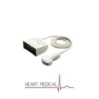
| C5-1 | General imaging, deep abdominal scanning | Abdomen, OB/GYN, Fetal Echo, Pediatric, Vascular | Curved Array | 1-5 MHz | Wide (111°) |
| C8-5 | Neonatal and pediatric imaging | Neonatal head, pediatric abdomen, vascular | Micro-Convex | 5-8 MHz | Compact (122°) |
| C9-2 | Focused imaging in OB/GYN | OB, GYN, Fetal Echo, Pediatric abdomen | Micro-Convex | 2-9 MHz | 102° |
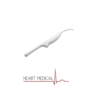
| C10-3v | Endocavitary applications | OB, GYN, Urology, Fetal Echo | Endovaginal | 3-10 MHz | 163° |
| C10-4EC | OB/GYN and urology exams | OB, GYN, Urology, Fetal Echo | Endocavitary | 4-10 MHz | 147° |
| D2cwc | Adult cardiac Doppler assessments | Adult Echo | CW Doppler | 2 MHz | N/A |
| D2tcd | Transcranial Doppler (TCD) | Neurological Doppler | CW Doppler | 2 MHz | N/A |
| D5cwc | Peripheral vascular Doppler studies | Vascular | CW Doppler | 5 MHz | N/A |
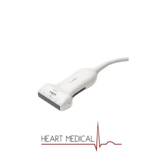
| L12-3 | Multi-purpose imaging with high detail | Vascular, Musculoskeletal, Small Parts, Abdomen | Linear | 3-12 MHz | 38 mm |
| L12-5 50 | Larger field-of-view vascular imaging | Pediatric, Musculoskeletal, OB, Small Parts | Linear | 5-12 MHz | 50 mm |
| L15-7io | Intraoperative procedures | Epiaortic, Musculoskeletal, Vascular | High-Frequency Linear | 7-15 MHz | N/A |
| L18-5 | High-resolution small structure imaging | Small Parts, Vascular, Pediatric, Musculoskeletal | Linear | 5-18 MHz | 38.9 mm |
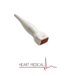
| S5-1 | Cardiac and TCD imaging | Adult Echo, Pediatric, Transcranial Doppler | Sector Array | 1-5 MHz | 20.3 mm |
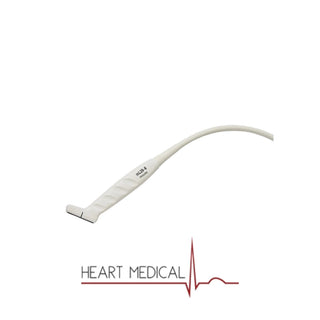
| S7-3t | Pediatric and adult transesophageal echocardiography (TEE) | TEE, Pediatric Echo | Transesophageal | 3-7 MHz | 5 mm |
| S8-3 | High-resolution adult echocardiography | Adult Echo, Pediatric | Sector Array | 3-8 MHz | 15.4 mm |
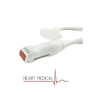
| S12-4 | Neonatal and pediatric cardiac imaging | Neonatal Echo, Pediatric Cardiac | Sector Array | 4-12 MHz | 9.78 mm |
| V6-2 | OB and fetal imaging with volume rendering | OB, Fetal Echo | 3D/4D Volume | 2-6 MHz | 100° |
| X5-1 | High-performance cardiac imaging with real-time 3D | Adult Echo, Vascular | xMATRIX Array | 1-5 MHz | 100° |
| X6-1 | High-resolution OB and general imaging | OB, Fetal Echo, GYN, Interventional | xMATRIX Array | 1-6 MHz | 100° |
| X7-2t | 3D transesophageal cardiac imaging | Adult Echo (TEE) | 3D TEE Probe | 2-7 MHz | N/A |
| X8-2t | Next-generation 3D transesophageal cardiac imaging | Advanced TEE | 3D TEE Probe | 2-7 MHz | N/A |
| VL13-5 | Breast and small part imaging with high resolution | Breast, Small Parts, OB, Vascular | Linear | 5-13 MHz | 38.4 mm |
| eL18-4 | Premium high-frequency imaging for detailed diagnostics | Breast, Small Parts, Musculoskeletal, Pediatric, Vascular | Linear | 2-22 MHz | N/A |
Role of Transducers in Ultrasound Imaging
Transducers (or probes) are the "camera" of the ultrasound system. They:
- Send sound waves into the body.
- Capture echoes to create images.
- Critical for accuracy: The right transducer ensures sharp images and reliable diagnoses.
Fundamentals of Ultrasound Transducers
Basic Principles
- Piezoelectric Effect: Crystals inside the transducer convert electricity into sound waves (and vice versa).
- Frequency:
- High frequency (e.g., 12 MHz): Better resolution for shallow structures (e.g., veins).
- Low frequency (e.g., 2 MHz): Deeper penetration for organs like the liver or heart.
- Trade-off: Higher frequency = sharper images but less depth; lower frequency = deeper view but fuzzier details.
Key Components
|
Category |
Component |
Description |
|
Key Components |
Crystal Arrays |
Generate and receive sound waves. |
|
Key Components |
Matching Layer |
Helps sound waves enter the body smoothly. |
|
Key Components |
Backing Material |
Reduces unwanted vibrations for clearer images. |
|
Philips Innovations |
PureWave Technology |
Uses advanced crystals for better image quality in tough cases (e.g., obese patients). |
Overview of Philips Epiq 5/7 Transducers
Standard Probes:
|
Probe |
Frequency |
Best For |
|
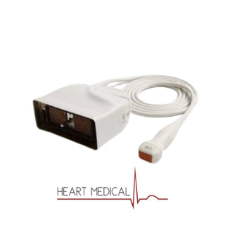
X5-1 PureWave |
1–5 MHz |
Adult heart, abdominal, and brain imaging. |
|
X6-1 PureWave |
1–6 MHz |
Pediatric heart and abdominal scans. |
|
S5-1 PureWave |
1–5 MHz |
General imaging (e.g., pregnancy, abdomen). |
|
L12-5 High-Density |
5–12 MHz |
Veins, arteries, tendons, and small organs. |
|
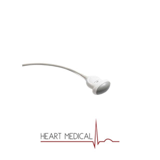
C9-2 Curved Array |
2–9 MHz |
Wide abdominal, OB/GYN, and bladder scans. |
Specialty Probes:
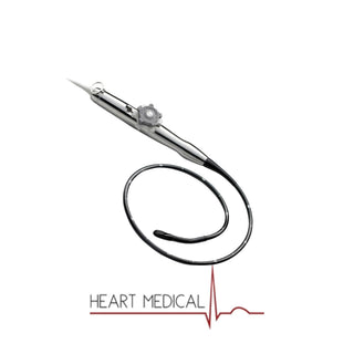
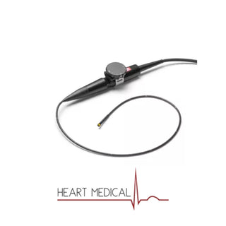
- TEE Probes (e.g., X7-2t): Inserted into the esophagus for detailed heart imaging.
- 4D Matrix Probes (e.g., X8-2t): Captures live 3D videos of babies in the womb.
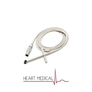
Probe Guides: Function and Integration
A probe guide is a clip-on tool that helps doctors steer needles during procedures like biopsies or injections.
- Uses: Liver biopsies, draining fluid, or placing IVs.
- Types: Disposable (sterile) or reusable.
Using the Probe Guide:
- Attach: Clip the guide onto the transducer.
- Align: Line up the guide’s markings with the ultrasound screen’s needle path.
- Adjust: Tilt the probe to hit the target safely.
- Safety: Always sterilize reusable guides and check alignment before use.
Operation and Image Optimization
Imaging Modes & Technologies:
- B-mode: Standard grayscale imaging.
- Doppler: Shows blood flow (red/blue colors).
- 3D/4D: 3D snapshots or live 3D video (e.g., baby’s face).
- Elastography: Measures tissue stiffness (helps detect tumors).
Philips’ Advanced Tools:
- Beamforming: Sharpens images by focusing sound waves.
- Auto-Optimize: Press a button to let the system adjust settings for you.
Optimizing Image Quality:
- Adjust Depth: Zoom in/out to focus on the area of interest.
- Tweak Gain: Brighten or dim the image.
- Use Harmonics: Reduces noise for cleaner images.
- Pro Tip: Apply enough gel and press the probe gently to avoid blurry images.
Workflow Integration Tips
Switching Probes During an Exam:
- Example: Start with the C9-2 curved array for a general abdominal scan, then switch to the L12-5 for detailed liver vessel imaging.
- Save Time: Use the system’s preset organ settings (e.g., "Liver" or "Carotid Artery") to auto-adjust depth and frequency.
Saving and Exporting Images:
- 4D Obstetric Scans: Save short video clips to track fetal movement.
- Needle Guidance: Capture split-screen images showing the needle path and target area for procedure documentation.
Safety Considerations
Patient Safety:
- TEE Probes: Ensure proper sedation and monitor vital signs during esophageal insertion.
- Needle Guides: Double-check alignment to avoid accidental punctures of non-target areas.
Transducer Safety:
- Avoid Mismatches: Using a high-frequency probe (e.g., L12-5) for deep abdominal scans may miss critical details—stick to low-frequency probes like the C9-2.
- Thermal Safety: Never leave a probe stationary on the skin for long periods during Doppler exams.
Selecting the Right Transducer
Clinical Considerations:
- Heart: Use X5-1 or X6-1 (phased array for tight spaces between ribs).
- Baby Scans: C9-2 (curved array for wide womb views).
- Veins: L12-5 (high frequency for crisp details).
Maintenance and Care
- Cleaning: Wipe probes with approved disinfectant wipes after each use.
- Storage: Hang probes to avoid cable damage; don’t bend connectors.
- Troubleshooting:
- Image Dropout: Check for damaged cables or loose connections.
- Blurry Images: Reapply gel or adjust depth/gain.
Conclusion
The Philips Epiq 5/7, with its advanced transducers and intuitive tools, is a powerhouse for modern diagnostic imaging. By:
- Choosing the right probe (e.g., X5-1 for cardiac scans).
- Using probe guides safely for precise needle procedures.
- Optimizing settings like depth and gain for crystal-clear images.
You can enhance diagnostic accuracy and patient care. Whether you’re guiding a biopsy or capturing a baby’s first 3D snapshot, the Epiq 5/7 adapts to your needs, proving why it’s a trusted tool in clinics worldwide.
Frequently Asked Questions
1. What makes the Philips EPIQ 5/7 different from other ultrasound systems?
Unlike standard ultrasound machines, the EPIQ 5/7 features PureWave Crystal Technology for superior image clarity, even in patients with high BMI. It also integrates Anatomical Intelligence, which automatically adjusts settings for specific organs, reducing the need for manual fine-tuning.
2. Can EPIQ 5/7 transducers be used across multiple imaging applications?
Yes, many transducers designed for the EPIQ 5/7 are multi-purpose. For example, the L12-5 probe is used for vascular, musculoskeletal, and small parts imaging, while the X5-1 is optimized for both cardiac and general abdominal scans. However, specialized probes like the X7-2t (TEE) are designed for specific applications and should not be used interchangeably.
3. How does the EPIQ 5/7 handle challenging imaging conditions?
The system excels in difficult imaging scenarios due to PureWave technology, which enhances signal penetration, making it particularly useful for obese patients or deep organ scanning. Additionally, adaptive beamforming helps improve image contrast and detail resolution in complex cases.
4. What factors should be considered when selecting a transducer for the EPIQ 5/7?
When choosing a transducer, consider:
- Depth vs. Resolution – Lower frequencies (e.g., C5-1, 1-5 MHz) penetrate deeper, while higher frequencies (e.g., L18-5, 5-18 MHz) provide finer detail for shallow structures.
- Field of View – Probes like C9-2 have a wide field of view, making them ideal for obstetric and abdominal imaging.
- Specialty Applications – TEE probes (X7-2t, X8-2t) are exclusively designed for transesophageal cardiac imaging and are not suitable for other exams.
5. How does Philips' 3D/4D technology improve diagnostic capabilities?
Philips offers real-time 3D and 4D imaging with xMATRIX probes (X5-1, X6-1, X8-2t), which allow for:
- Live fetal scans with detailed anatomy views.
- 3D cardiac imaging for better visualization of heart defects.
- Guided interventions, reducing reliance on multiple 2D image slices.
6. What are the best practices for maintaining and cleaning EPIQ 5/7 transducers?
To ensure longevity and accurate imaging:
- Use approved disinfectants – Avoid alcohol-based wipes for certain probes, especially TEE transducers.
- Store properly – Hang transducers instead of coiling cables to prevent damage.
- Check connections – Loose or damaged cables can cause image artifacts or dropouts.
7. Can EPIQ 5/7 transducers be used with older Philips ultrasound systems?
Most transducers are system-specific, meaning not all EPIQ 5/7 probes will be backward compatible with older Philips models. However, some shared transducers, like the C5-1 or S5-1, may work on certain Affiniti systems. Always check the Philips compatibility chart before attempting cross-system use.
Why Choose Heart Medical for Your Philips Ultrasound Needs?
At Heart Medical, we are authorized resellers of Philips ultrasound machines and transducers, providing new, demo, and refurbished systems with nationwide delivery.
- Genuine Philips Equipment
- Nationwide Delivery
- Expert Support
- Cost-Effective Solutions
Get in Touch Today
Reviewed by Heart Medical Clinical Applications Team
Clinical and technical specialists ensuring accuracy and relevance across all Heart Medical content.
Related Products