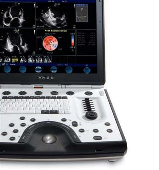
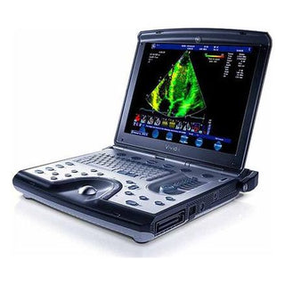
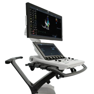
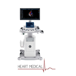
The GE Vivid q is a portable cardiovascular ultrasound system designed to bring high-end imaging and advanced quantification tools into a compact, mobile platform. Built for cardiologists, vascular specialists, and clinicians who need accuracy on the move, the Vivid q delivers premium image quality with features like Tissue Velocity Imaging (TVI), Automated Function Imaging (AFI), and Tissue Synchronization Imaging (TSI). Whether in a hospital, cath lab, or mobile clinic, this system combines portability with performance, making it one of GE’s most versatile cardiovascular solutions.
GE Vivid q Features

1: System Architecture
- High-performance, battery-operated, ultra-portable diagnostic ultrasound system.
- Based on GE’s TruScan architecture (used across GE cardiovascular ultrasound family) with raw data storage and advanced post-processing.
2: Display / Monitor & Physical Design
- 15″ high resolution TFT LCD monitor.
- Display resolution 1600×1200 pixels, supporting ~260,000 simultaneous colors.
- Screen can tilt / close from 0-180°, wide viewing angles. When closed, screen auto enters standby and reboots quickly when opened.
- Brightness, contrast, blue-tint adjustment for different ambient light environments.
3: Data Acquisition & Probes
- Supports multiple probe types: Phased Array, Linear, Curved (convex), TEE, 2D Intracardiac Echo (ICE), etc.
- M4S-RS matrix array transducer (for adult cardiac) gives improved image quality and deeper penetration.
- Wide aperture mode for convex & linear probes, which improves penetration and spatial resolution for deeper structures.
4: Imaging Modes & Processing
- 2D imaging, harmonic tissue imaging, coded octave imaging.
- Doppler modes: Color Doppler, Pulsed Wave (PW), Continuous Wave (CW), Power Doppler, etc.
- Tissue Doppler (Velocity), Tissue Velocity Imaging (TVI) for assessing motion.
- Tissue Tracking (TT), Tissue Synchronization Imaging (TSI) – to assess mechanical synchronicity of myocardial segments.
- Automated Function Imaging (AFI) – for left ventricle strain / strain rate, global/regional function quantification.
5: Image Quality Enhancements & Workflow Tools
- Speckle Reduction (SRI HD) to reduce artifact and improve clarity.
- Smart Depth – automatically optimize imaging parameters depending on scan depth.
- Automatic Tissue Optimization (ATO) of real-time 2D images.
- Variable transmit frequencies, contour filtering, dynamic range, zoom functionality, grayscale parameter control (gain, reject etc) per depth.
6: Display & Review Features
- Instant-review screen: displays 12 simultaneous loops/images to review study quickly.
- Display configurations: single, dual, quad, split-screen, duplex, triplex (side-by-side, top-bottom etc) for live, replay and clipboard.
7: Depth & Penetration
- Max imaging depth up to ~30 cm (depending on probe) for deeper structures.
8: Power / Portability / Battery
- Rechargeable battery option; power-efficient design. Over one hour scanning on battery in some configurations.
- When in standby (screen closed) battery still supports quick boot-up from standby.
9: Connectivity & Data Storage
- Raw data storage for offline post-processing, measurements.
- Ability to export via DICOM, network, USB, CD/DVD, external hard drives.
- Compliance with DICOM standards (image storage, multi-frame storage, query/retrieve, storage commitment etc) for integration into PACS etc.
10: Annotations, Display Overlays & User Interface
- Annotations like Mechanical Index (MI), Thermal Index, patient name & ID, hospital name, time/date, probe name, scanning parameters etc.
- Multi‐language support for UI, reports and manual.
11: Extras / Special Features
- Contrast imaging support (for abdominal contrast, etc) with coded phase inversion in certain probes.
- IMT (Intima-Media Thickness) analysis package for vascular-risk early detection.
- B-Flow / Blood Flow Imaging (BFI) for cleaner flow display without color artifacts.
Probe Guide
| Probe Type | Model | Frequency Range | Best Applications |
|---|---|---|---|
| Phased Array (Sector) | M4S-RS | 1.5 – 3.6 MHz | Adult cardiac; deep penetration with strong overall image quality |
| Phased Array (Sector) | 5S-RS | 2.0 – 5.0 MHz | Pediatric cardiac; fetal heart; balanced depth + resolution |
| Phased Array (Sector) | 6S-RS | 2.7 – 8.0 MHz | Neonatal/pediatric cardiac; high-resolution shallow cardiac |
| Phased Array (Sector) | 7S-RS | 3.5 – 8.0 MHz | High-frequency sector imaging; fine wall/structure detail |
| Phased Array (Sector) | 10S-RS | 4.0 – 11.5 MHz | Very high-resolution shallow cardiac; pediatrics |
| Linear Array | 8L-RS | ~4.0 – 13.0 MHz | Vascular; small parts; MSK; superficial structures |
| Linear Array | 12L-RS | ~4.0 – 13.0 MHz | High-detail superficial imaging; vascular; small parts |
| Convex / Curved Array | 4C-RS | ~1.8 – 6.0 MHz | Abdominal; deeper organs; general curved imaging |
| Convex / Curved Array | 8C-RS | ~4.7 – 11.0 MHz | Shallow abdominal/superficial curved imaging |
| Intra-operative / Specialty | i12L-RS (hockey-stick) | ~5.0 – 13.0 MHz | Intra-operative; small organ/surgical access; superficial |
| TEE (Transesophageal) | 6T-RS / 6Tc-RS | ~2.9 – 8.0 MHz | Transesophageal cardiac imaging; peri-/intra-operative echo |
| TEE (Pediatric/Neonatal) | 9T-RS | ~3.0 – 10.0 MHz | Pediatric/neonatal TEE; higher-frequency esophageal imaging |
| Pencil / Doppler | P2D (≈2 MHz) | ~2.0 MHz | Spectral Doppler; flow velocity in deeper vessels |
| Pencil / Doppler | P6D (≈6 MHz) | ~6.0 MHz | Spectral Doppler; higher-frequency superficial flow |
| ICE Catheters* | AcuNav 8F/10F; SoundStar variants | ~4.5 – 11.5 MHz (variant-dependent) | Intracardiac echo for EP procedures |
Clinical Applications
The GE Vivid q is engineered for a wide range of cardiovascular and general imaging applications, making it a versatile system for hospitals, clinics, and mobile care environments.
- Cardiac Imaging – Advanced quantification tools like Automated Function Imaging (AFI), Tissue Velocity Imaging (TVI), and Tissue Synchronization Imaging (TSI) provide precise assessment of ventricular function, wall motion, and myocardial mechanics.
- Vascular Studies – IMT (Intima-Media Thickness) package and high-frequency linear probes support detailed carotid and peripheral vascular assessments.
- Stress Echocardiography – Supports exercise and pharmacological stress testing with real-time wall motion analysis.
- Transesophageal Echocardiography (TEE) – Compatible with adult and pediatric TEE probes for intra-operative and peri-operative cardiac evaluation.
- Pediatric & Neonatal Cardiology – Small footprint and high-frequency transducers allow safe, detailed imaging for pediatric and neonatal patients.
- Intra-cardiac Echocardiography (ICE) – Provides clear visualization during electrophysiology procedures.
- Abdominal & Small Parts Imaging – Convex and linear probes extend use to abdominal, musculoskeletal, and superficial structures when required.
- Contrast Imaging – Coded phase inversion technology enhances blood flow visualization and improves cardiac perfusion assessment.
Advantages vs Limitations
Advantages
- High-Quality Cardiac Imaging – Advanced quantification tools like AFI, TVI, and TSI give detailed functional assessment for cardiology.
- Portability – Lightweight, battery-powered laptop design enables use in mobile clinics, cath labs, and bedside environments.
- Probe Versatility – Wide range of transducers including sector, linear, convex, TEE, and ICE catheters for diverse clinical needs.
- Workflow Efficiency – Smart Depth, Speckle Reduction (SRI HD), customizable presets, and raw data post-processing save time and improve consistency.
- Connectivity – DICOM, USB, and DVD support allow easy data sharing across hospital systems and PACS.
- Broad Clinical Applications – Suitable for adult and pediatric cardiac imaging, vascular studies, abdominal scans, and intra-operative use.
Limitations
- Aging Hardware – Originally launched more than a decade ago, it lacks some of the newer AI-driven features found in latest GE systems.
- Battery Duration – While portable, extended scanning on battery is limited compared to newer portable ultrasounds.
- Learning Curve – Advanced quantification tools require training for accurate use.
- Cost of Probes – Specialized probes (like TEE or ICE) can be expensive, and availability may vary depending on region.
- System Age / Support – Being an older model, software updates and OEM support may be limited compared to current GE platforms.
GE Vivid q vs Other GE Ultrasound Systems
| Feature | GE Vivid i | GE Vivid q | GE Vivid E9 |
|---|---|---|---|
| Portability (Laptop Design) | ✔ | ✔ | ✘ (Cart-Based) |
| Advanced Quantification (AFI, TSI, TT) | ✘ | ✔ | ✔ |
| Speckle Reduction Imaging (SRI HD) | ✘ | ✔ | ✔ |
| Matrix Array Probe (M4S-RS) | ✘ | ✔ | ✔ |
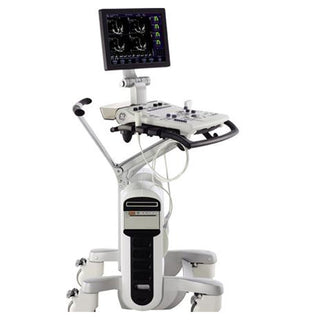
| 4D/Real-Time 3D Cardiac Imaging | ✘ | ✘ | ✔ |
| Battery Operation | ✔ | ✔ | ✘ |
| Touchscreen Interface | ✘ | ✘ | ✔ |
| Wide Probe Compatibility (TEE, ICE, Linear, Convex) | Limited | ✔ | ✔ |
| Raw Data Storage / Post-Processing | ✔ (Basic) | ✔ (Enhanced) | ✔ (Full) |
| Target Environment | Portable / Mobile | Portable + Advanced Cardiac | Premium Hospital / Cath Lab |
Conclusion
The GE Vivid q remains one of GE Healthcare’s most trusted portable cardiovascular ultrasound systems. With its advanced quantification tools, versatile probe compatibility, and reliable portability, it continues to support cardiologists, vascular specialists, and clinicians who need dependable imaging at the bedside or in mobile care. While newer models like the Vivid iq bring updated designs, the Vivid q offers an excellent balance of performance, portability, and value for a wide range of cardiovascular applications.
Looking for a GE Vivid q ultrasound system or compatible probes?
Heart Medical provides genuine equipment, replacement parts, and service support with worldwide shipping and expert assistance.
Request a Quote today and equip your clinic with a proven cardiovascular imaging solution.
Reviewed by Heart Medical Clinical Applications Team
Clinical and technical specialists ensuring accuracy and relevance across all Heart Medical content.