This review is based on official data sheets and user manuals. For specific quotes and inquiries about the ultrasound system, please contact Heart Medical team.
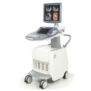
Overview of GE Vivid E95 Ultrasound Machine
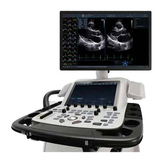
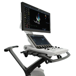
The GE Vivid E95 ultrasound system is a premium 4D cardiovascular ultrasound machine designed to address some of the most complex challenges in modern healthcare. The system is particularly well-suited for cardiovascular imaging and offers detailed insights into cardiac structures and function, which are critical for accurate diagnosis and treatment planning. At the heart of the Vivid E95 is the cSound® Technology, a software-based beamformer image reconstruction platform. This technology allows the system to get more information upfront, which enables superior image quality and faster processing times.
It has advanced imaging capabilities, including 2D, 3D, and Doppler modalities. The machine has fast HD algorithms that enhance image clarity and resolution. It has an ergonomic design that provides easy usage and helps in maintaining a great workflow during examinations. The Vivid E95 also supports various transducer options that meet patient needs and clinical scenarios. Let’s get to know about the list of features, specifications, and usage in detail further
Key Features of GE Vivid E95 Ultrasound System
Visualization
- Exceptional 2D and 4D Image Quality
The Vivid E95 delivers uncompromised image quality. It achieves high contrast and spatial resolution which enables detailed imaging crucial for accurate diagnoses. This is particularly beneficial in challenging cases, such as those involving obese or difficult-to-scan patients. This system is integrated with advanced imaging technology such as 2D and 3D which provide detailed cardiac assessment. The cSound Architecture contributes to exceptional image uniformity and clarity.
- XDclear® Probe Technology: Captures data from every channel in the probe
XDclear® probe technology maximizes data capture, enhancing near-field resolution, lateral wall definition, and image uniformity. This ensures consistent performance across various applications, including cardiac, vascular, and abdominal imaging. This technology provides excellent color sensitivity, contrast resolution, and spectral crispness.
- True Confocal Imaging (TCI)
True Confocal Imaging (TCI) provides uniform focusing throughout the field of view, combined with increased contrast resolution. This maintains high image quality at high frame rates, which is valuable during procedures like TEE.
- HDlive™
HDlive™ is an advanced visualization technique that creates lifelike, photorealistic representations of anatomical structures which enables easier and more accurate interpretation. FlexiLight also contributes to photo-realistic imaging by allowing the positioning of light sources.
- Triplane Imaging:
Triplane Imaging allows clinicians to image three planes from a single heartbeat. This provides comprehensive cardiac assessments with high temporal and spatial resolution, which helps in reducing exam time.
- Virtual Apex:
Phased array probes provide a wide field of view with the virtual apex feature. This feature enhances the visualization of structures located at the edges of the imaging sector and ensures that the full extent of cardiac anatomy can be captured.
Quantification
These features allow for precise measurements and assessments of cardiac function. Here's a breakdown:
- Automated Function Imaging (AFI)
This tool automatically analyzes the movement of the left ventricular walls while the heart is at rest. It goes beyond visual assessment by calculating a range of numerical values (parameters) that represent how well the heart is pumping. Crucially, it includes "AFI Stress" protocols. This means it can also quantify how the heart's walls move during stress echocardiography. This allows doctors to detect subtle issues that might only appear under stress. This is useful for detecting coronary artery disease.
- 4D Strain
"Strain" refers to the deformation or stretching of the heart muscle. This feature uses an algorithm to track the movement of "speckles" within the heart tissue in 4D (three dimensions plus time). This is valuable for detecting early signs of heart disease that might not be visible with traditional imaging.
- 4D Auto AVQ (Aortic Valve Quantification)
This feature automates the process of measuring the aortic outflow tract. It automatically segments (outlines), aligns, and quantifies this area. This is extremely important for planning and performing TAVI/TAVR procedures, where a new valve is inserted through a catheter.
- 4D Auto MVQ (Mitral Valve Quantification)
Similar to AVQ, this feature focuses on the mitral valve. It uses a semi-automatic algorithm to detect the surface of the mitral valve, which provides detailed visualization and quantification. It supports transesophageal echocardiography (TEE) images, which provide very clear views of the heart.
Workflow Enhancements
- Streamlined Exams
The Vivid E95 is capable of making complex 4D imaging as straightforward as standard 2D imaging. This is achieved through user-friendly tools and an intuitive interface. The system's DICOM compatibility adapts to user preference to ensure optimal image display.
Features like "QuickAgger" and "Auto-Doppler" automate routine tasks which reduces the need for manual adjustments and speed up exams.
- 4D TEE (Transesophageal Echocardiography)
The Vivid E95 simplifies and accelerates 4D TEE procedures to provide clear and precise images. Powered by cSound, the system delivers high-quality images that are crucial for accurate assessment and diagnosis during TEE.
- Multi-slice Imaging
This feature allows clinicians to extract standard long-axis and short-axis views from 4D volume datasets. This can be done in real-time (live mode) or from previously recorded data (replay mode).
- Laser Lines
Laser Lines improve spatial awareness, especially during complex imaging tasks that make it easier to understand the 3D structure of the heart.
Advanced Technologies
The GE Vivid E95 is equipped with several advanced technologies that enhance its imaging capabilities, streamline workflows, and improve diagnostic accuracy. Here is an overview of the technologies:
- cSound® 2.0
This software makes super-detailed 4D heart images in just one heartbeat. This reduces the time needed for image acquisition, especially in patients with irregular heart rhythms. It also eliminates artifacts caused by heart rate variability to provide clearer and more accurate images.
- FlexiSlice
This feature allows doctors to slice and view 4D heart images in 2D or 3D, either live or after the scan. This reduces the need for multiple scans by allowing users to manipulate and extract views from a single 4D image.
- FlexiViews
It provides pre-set views of the heart during live imaging. By offering predefined views, FlexiViews minimizes the time needed to set up and adjust imaging parameters during the procedure. This simplifies the imaging process, allowing clinicians to focus on the procedure rather than adjusting the system.
- CT Fusion
It combines 4D ultrasound images with CT scans on the same screen. This helps doctors see the heart in more detail by merging two types of images which reduces the need for multiple imaging modalities that save time and improve workflow efficiency
Security and Connectivity
The GE Vivid E95 is designed to keep patient data safe and make it easy to share information with other healthcare systems. Here’s how it works:
- LDAP (Lightweight Directory Access Protocol)
This feature controls who can access the system. So, only authorized users can log in to keep patient data secure. This reduces the risk of data breaches.
- Disk Encryption
The Vivid E95 protects patient data stored on the system. So, even if the system is stolen, the data stays safe and private. This ensures compliance with privacy regulations.
- Windows® 10 Operating System
It uses application whitelisting to block unauthorized programs. This prevents harmful or unauthorized software from running on the system.
- DICOM Support
It allows the system to share images and data with other medical systems. This provides enhanced support for cardiac and vascular DICOM SR ensuring accurate and fast sharing of heart-related data. It also makes it easy to integrate with hospital networks and electronic medical records (EMR).
- Tricefy® Uplink™
It quickly uploads images and patient data to the Tricefy Cloud and enables easy sharing of images with other doctors or patients. This also provides a long-term storage solution for patient data.
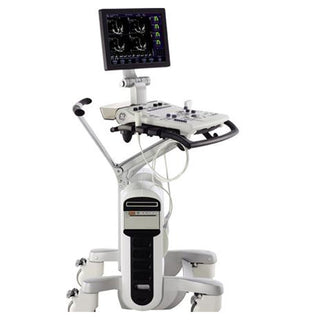
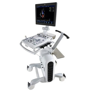
Ergonomics and User Experience
Here is how the GE Vivid E95 makes the user experience comfortable, efficient, and easy:
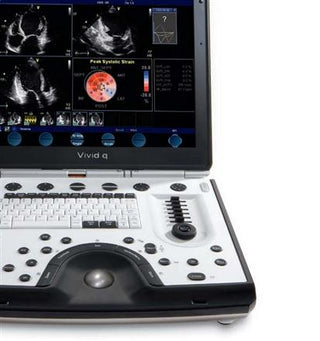
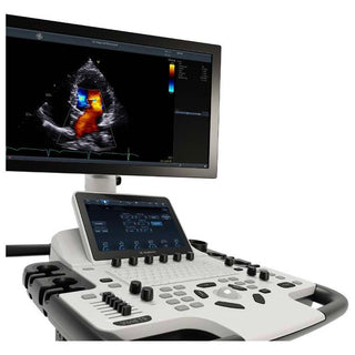
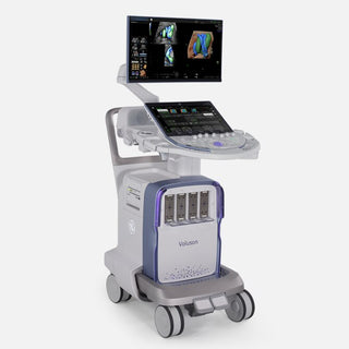
- 22" High-Resolution OLED Monitor
It displays super-clear, high-quality images and helps doctors see even the smallest details in heart scans.
- 12" LCD Touch Screen
This acts like a tablet for easy navigation and makes it simple to adjust settings and view patient data.
- Adjustable Floating Keyboard
It can be moved to the most comfortable position which reduces strain during long scanning sessions.
- Easy Mobility
The system is designed to be moved around easily which makes it flexible for different clinical settings.
- Low Power Consumption
It uses less energy which reduces operating costs.
- User-Friendly Interface
The configurable touch panel allows users to customize the interface to suit their preferences. The Image Manager provides quick access to patient images and data.
| Specification | Details |
|---|---|
| Display | Choice of a 22" high-resolution OLED monitor or a 24" high-contrast LCD monitor. Additionally, equipped with a 12" high-resolution LCD multi-touch screen. [Source] |
| Weight | Approximately 128 kg (283 lbs). [Source] |
| Dimensions | Height: 141 cm (55.5 in); Width: 58.5 cm (23 in); Depth: 83 cm (32.7 in). [Source] |
| Transducer Frequency | Supports a range of transducers with frequencies from 1 to 12 MHz, including single crystal probes and advanced 4D multiplane cardiac and TEE imaging. [Source] |
| Imaging Modes | 2D, 3D/4D imaging, including advanced modes like HDlive™, Triplane imaging, and Virtual Apex. [Source] |
| Battery Life | Not specified; the system is typically powered via an AC connection. |
| Connectivity | Ethernet, USB, and optional wireless adapter. [Source] |
| Power Source | AC connection with voltage ranging from 100-240 VAC ±10% and power consumption of 700W / 770 VA. [Source] |
| Storage Capacity | Equipped with a 500GB hard drive for data storage. [Source] |
| Operating System | Windows 10. [Source] |
| Optional Features | Advanced quantification tools such as Automated Function Imaging (AFI), 4D Auto AVQ, 4D Strain, and 4D Stress Echo. [Source] |
Service and Support
The GE Vivid E95 comes with excellent service and support options to keep the system running smoothly and help healthcare providers focus on patient care. Here’s what’s available:
Assurepoint® Services
This reduces downtime so that the system is always available when needed. Proactive monitoring and remote support prevent problems before they happen.
Iking®
GE provides direct access to technical experts. Doctors and technicians can get quick answers to technical questions, often in just a few minutes.
InSite®
GE specialists can troubleshoot and fix issues remotely, often without needing to visit the hospital. This minimizes downtime and keeps the system operating at peak performance.
QA Care
GE provides documentation support for ultrasound accreditation. This helps hospitals meet regulatory requirements for image quality and system performance/
How to Use Vivid E95
System Preparation
- Power Connection: Connect the ultrasound machine to a grounded AC power source using the provided power cable. Ensure the system's voltage setting matches the available mains voltage to prevent equipment damage.
- System Activation: Press the power button located on the control panel to turn on the system.
- Software Verification: Verify that the system software is up-to-date. Access the software version information through the system's settings menu.
Patient Preparation
- Medical History Review: Review the patient's medical records to understand the clinical context and determine the appropriate imaging protocol.
- Procedure Explanation: Clearly explain the ultrasound procedure to the patient to ensure they are comfortable and informed.
- Patient Positioning: Position the patient according to the specific examination requirements, ensuring optimal access to the area of interest.
Transducer Selection and Application
- Transducer Choice: Select the appropriate transducer based on the examination type and patient characteristics. The Vivid E95 supports a range of transducers with frequencies from 1 to 12 MHz.
- Transducer Inspection: Before use, inspect the transducer for any signs of damage or wear. Do not use a damaged transducer.
- Gel Application: Apply a sufficient amount of ultrasound gel to the transducer to ensure proper acoustic coupling.
Imaging Procedure
- Mode Selection: Choose the appropriate imaging mode (e.g., 2D, 3D/4D, Doppler) based on the clinical requirements.
- Parameter Adjustment: Adjust imaging parameters such as gain, depth, and focus to optimize image quality. Utilize the system's automatic optimization features when available.
- Doppler Assessment: If evaluating blood flow, switch to the Doppler mode and adjust the sample volume and angle to obtain accurate measurements.
- Continuous Monitoring: Continuously monitor the real-time images, making necessary adjustments to maintain optimal visualization.
Image Documentation
- Image Capture: Use the system's capture function to record essential images and measurements.
- Data Storage: Save the captured data to the system's internal storage or export it to an external device as needed.
- Data Labeling: Accurately label all images and measurements with patient information and relevant annotations.
Post-Examination Procedures
- Data Integration: Transfer the saved data to the Electronic Medical Record (EMR) system, ensuring compliance with institutional protocols.
- Transducer Cleaning: Thoroughly clean the transducer and remove any residual gel to prevent cross-contamination. Follow the manufacturer's guidelines for cleaning and disinfection.
- System Shutdown: Properly shut down the ultrasound system following the manufacturer's recommended procedures.
Major Applications of the Vivid E95
The GE Vivid E95 Ultrasound System is a versatile imaging tool designed for a wide range of clinical applications with a focus on cardiovascular imaging. Here are the major applications of the Vivid E95:
Cardiovascular Imaging
2D and 4D Echocardiography: Provides high-resolution images of the heart’s structures, including chambers, valves, and walls.
Transesophageal Echocardiography (TEE)
4D TEE Imaging offers detailed, real-time 3D and 4D views of the heart during TEE procedures. Single-Beat Acquisition captures high-quality 4D images in a single heartbeat, even in patients with irregular heart rhythms.
Stress Echocardiography
AFI Stress Protocols quantify left ventricular wall motion at rest and during stress (exercise or pharmacological). Segmental and Global Strain Analysis assesses heart function under stress, helping to diagnose ischemic heart disease and evaluate the effectiveness of treatments.
Pediatric and Congenital Heart Disease
Micro TEE Multiplane Probe is designed for neonatal and pediatric patients. This probe allows for detailed imaging of complex heart conditions in small patients (down to 2.5 kg).
The Vivid E95 provides detailed imaging for diagnosing and managing conditions like atrial septal defects (ASD), ventricular septal defects (VSD), and tetralogy of Fallot.
Interventional Cardiology
TAVI/TAVR Support provides precise imaging for planning and guiding transcatheter aortic valve implantation/replacement procedures.
Mitral Valve Interventions assist in diagnosing and planning treatments for mitral valve regurgitation or stenosis
Vascular Imaging
The Vivid E95 allows for carotid artery imaging to assess blood flow and detect blockages in the carotid arteries. It is also capable of performing peripheral vascular imaging to evaluate blood flow in arms and legs. Abdominal aortic imaging helps in detecting aneurysms or other abnormalities in the abdominal aorta.
Abdominal Imaging
This system is capable of performing Liver, Kidney, and Spleen Imaging. It also has applications in renal imaging, gallbladder and biliary tract imaging.
Obstetrics and Gynecology (OB/GYN)
This system is suitable for performing fetal imaging to monitor fetal development and detect abnormalities during pregnancy. It is also used in gynecological imaging to evaluate the uterus, ovaries, and other pelvic structures for conditions like fibroids or cysts.
Small-Parts Imaging
This system is capable of performing thyroid imaging, breast imaging and musculoskeletal imaging.
Vivid E95 Ultrasound Transducer Guide
| Probe Name | Frequency Range | Application |
|---|---|---|
| 6S-D | 1.0–5.0 MHz | Adult Cardiac Imaging |
| 4VC-D | 4.0–10.0 MHz | Pediatric and Adult Cardiac Imaging |
| 3Sc-RS | 1.7–3.4 MHz | 3D/4D Cardiac Imaging |
| 9L-D | 3.0–8.0 MHz | Vascular and Small Parts Imaging |
| C1-6-D | 1.0–6.0 MHz | Curved Array Abdominal Imaging |
| C2-9-D | 2.0–9.0 MHz | Curved Array Obstetrics Imaging |
| M5Sc-D | 1.5–4.6 MHz | General Imaging and Cardiac |
| 12S-D | 4.0–12.0 MHz | Musculoskeletal Imaging |
| 11L-D | 3.0–11.0 MHz | Linear Array Vascular Imaging |
| P2D | 2.0 MHz | Pencil Doppler Imaging |
| P6D | 6.3 MHz | Pencil Doppler Imaging |
| IC5-9D | 4.0–9.0 MHz | Rectal Imaging |
| 6Tc | 2.0–8.0 MHz | Transesophageal (TEE) Imaging |
| 6VT-D | 3.0–8.0 MHz | Transesophageal (TEE) Imaging |
Dos and Don’ts

Dos
- Make sure to position the probe accurately to get accurate imaging
- Always apply enough gel for clarity
- Make sure to know about each control to assess the data
- To save images during each assessment
- Make sure to update the software regularly
Don’ts
- Don’t speed up the scanning process
- Don't use a damaged transducer
- Don’t forget to use enough gel for clarity
- Don't skip routine maintenance and checks
Warranty Information
GE Healthcare typically offers the following for its premium ultrasound systems:
Standard Warranty
GE Healthcare usually provides a 1-year warranty covering parts and labor for manufacturing defects. This warranty ensures that any issues arising from normal use are addressed promptly at no additional cost.
Extended Warranty Options
GE often offers extended warranty plans that can be purchased to cover the system for additional years (e.g., 2–5 years). These plans may include preventive maintenance, software updates, and priority technical support
Conclusion
The GE Vivid E95 ultrasound machine is a powerful tool for cardiac imaging. This model is intended to have advanced features and capabilities that enhance diagnostic accuracy. Its user-friendly design and comprehensive functionality make it suitable for a wide range of clinical applications. By following the outlined best practices along with dos & don’ts and understanding its specifications, healthcare professionals can upgrade the benefits of this exceptional machine. They will get optimal assessment results for any clinical examination. For those who are looking to enhance their cardiac diagnostic capabilities, the GE Vivid E95 stands out as a leading and superior choice!
Explore more about GE Vivid E95 advanced ultrasound technology at Heart Medical and get this machine to support your advanced cardiac imaging needs.
Frequently Asked Questions
What makes the Vivid E95 different from other ultrasound systems?
The Vivid E95 stands out due to its ultra-high 4D imaging capabilities, advanced quantification tools, and streamlined workflows. It also offers single-beat acquisition for 4D imaging, making it faster and more efficient than many conventional systems.
Can the Vivid E95 be used for pediatric and neonatal imaging?
Yes, the Vivid E95 is equipped with a Micro TEE Multiplane Probe specifically designed for imaging complex heart conditions in neonatal patients (down to 2.5 kg) and adults with intolerance to standard TEE probes.
Can the Vivid E95 integrate with hospital systems?
Yes, the Vivid E95 supports DICOM for integration with hospital networks and electronic medical records (EMR). It also offers Tricefy® Uplink™ for cloud-based image sharing and storage
Reviewed by Heart Medical Clinical Applications Team
Clinical and technical specialists ensuring accuracy and relevance across all Heart Medical content.
Related Products