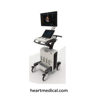
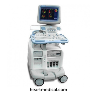
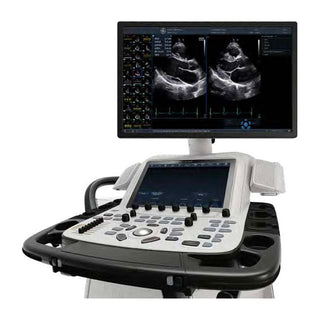
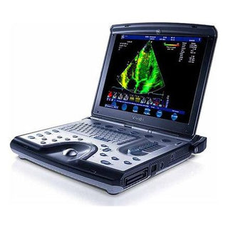
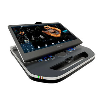
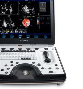
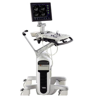
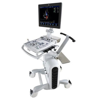
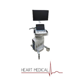
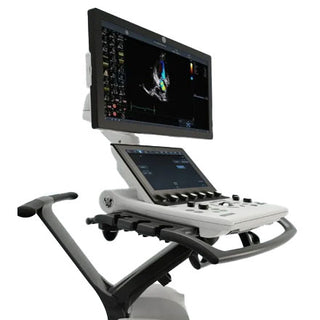
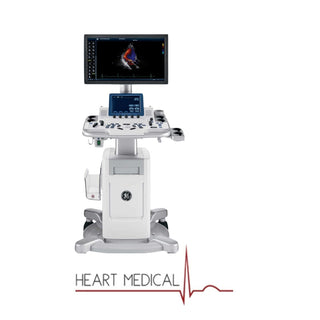
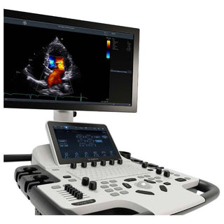
The GE Vivid S60 ultrasound system stands out for cardiovascular diagnostics. For healthcare facilities looking to acquire this machine without the high cost of a new unit, we recommend choosing a refurbished Vivid S60 ultrasound system in excellent condition. In this discussion, we will review the refurbished Vivid S60 in detail. Keep reading to find out if this system is suitable for your clinical practices.
Key Features of the GE Vivid S60 Ultrasound System

The GE Vivid S60 ultrasound system is a cardiovascular ultrasound machine that possesses advanced imaging capabilities with user-friendly features. Below are the key features Vivid S60 offers:
Image Quality and Processing
- cSound™ Software Beamformer: The cSound beamformer is the key component of the GE Vivid S60 ultrasound system that improves the quality of ultrasound images. Unlike traditional ultrasound systems that rely on hardware to form the beam, the Vivid S60 uses a software-based approach. This allows for more flexible and advanced processing of the data. The system captures data from all elements of the ultrasound probe and stores it temporarily in a "Local Big Data Memory." This ensures that no information is lost and allows for comprehensive analysis. The data is then processed by parallel processors, similar to those found in supercomputers. This enables the system to perform complex calculations quickly and efficiently, leading to improved image quality. The software-based approach and parallel processing power enable the system to generate clearer and more detailed images.
- Ultra Definition Clarity Filtering: Enhances image clarity and reduces speckle noise, which is that grainy texture you sometimes see in ultrasound images. By reducing speckles, the overall image clarity is improved which makes it easier to distinguish subtle differences in tissues.
- HD Imaging: Balances resolution, penetration, and image uniformity.
- Virtual Apex: This feature is specifically designed for phased array probes, which are commonly used in cardiac imaging. It expands the field of view, particularly in the near field (the area closest to the probe). This is helpful for visualizing structures that are wider than the probe itself, without having to move the probe around as much.
- Elevation Compound Imaging: Improves image quality by considering a wider slice for 2D imaging. This technique is particularly effective when using the 6VT probe, and enhances the overall image quality.
- Coded Phase Inversion: This technology focuses on improving the clarity of borders and enhancing contrast within the image. It reduces noise and strengthens the definition of tissue walls which makes it easier to see the boundaries between different structures.
- Multiple-Angle Compound Imaging: Combines images from different angles to enhance border definition and reduce angular dependence. This results in a more accurate and detailed representation of the underlying anatomy.
Advanced Imaging Modes
- 2D Imaging: Provides grayscale images of anatomical structures.
- Color Flow: Visualizes blood flow direction and velocity.
- Doppler: Measures blood flow velocity and patterns. The Vivid S60 offers high-quality versions of these basic modes with excellent spatial resolution.
- B-flow Imaging: It provides real-time images of blood reflectors (e.g., red blood cells), allowing for the visualization of vascular hemodynamics (blood movement) without the need for contrast agents.
- Tissue Velocity Imaging (TVI): Provides myocardial velocity data with color overlay. This helps in assessing heart function and identifying abnormalities.
- Strain and Strain Rate Imaging: Optional features for assessing myocardial deformation.
- Tissue Synchronization Imaging (TSI): Evaluates myocardial synchronicity (optional with Advanced QScan).
Ease of Use
- Touch Screen Control: The system features an intuitive touch screen interface, similar to a tablet or smartphone. This allows for direct interaction with the system's functions and settings. The touch screen also supports alpha-numeric keyboard entry for easy data input.
- Ergonomic FlexFit Design: The Vivid S60 is designed with user comfort in mind. You can adjust the height of the control panel to suit your preference. This reduces strain during long procedures. You can also easily position the monitor for optimal viewing.
- Automated Tools: This ultrasound system includes several automated tools to simplify workflows. The features include: 2D Auto EF which automates the measurement of Ejection Fraction (EF), a key indicator of heart function; AFI (Automated Function Imaging) Productivity Package, which assesses heart wall motion; and Scan Assist Pro which guides users through scanning procedures.
- Automated Image Optimization: The cSound technology automatically optimizes image quality which reduces the need for manual adjustments.
Portability
- Compact and Lightweight: Easy to transport with a fold-down monitor.
- Battery Option: Vivid S60 can be equipped with a battery that allows for quick setup and scanning at the patient's location, whether it's in a clinic, hospital room, or other setting. This eliminates the need to find a power outlet immediately.
Features to Perform Quantitative Analysis
- These features include Automated Ejection Fraction (AutoEF), Automated Function Imaging (AFI), and Intima-Media Thickness (IMT) Measurements which is an optional tool that automates the measurement of the Intima-Media Thickness (IMT) of the carotid artery.
Probe Technology
- XDclear™ Probes: Utilize Single Crystal, Acoustic Amplifier, and Cool Stack technologies for deep penetration, high sensitivity, and spatial resolution.
- Wideband Probes: The system supports a variety of probes to suit different types of exams. Common types include phased array, linear, convex, and TEE probes.
- Probe Presets: To simplify operation, the system includes pre-configured settings for various exam types. Presets streamline the setup process for common exams, such as cardiac, vascular, OB/GYN, and pediatric scans. These presets automatically adjust parameters like frequency, gain, and depth to optimize the system for the specific clinical application.
Workflow and Connectivity
- DICOM 3.0 Support: Enables easier integration with hospital networks for image storage and sharing.
- EchoPAC™ Integration: Advanced post-processing and analysis capabilities.
- InSite™ Express Connection: Remote service and training support.
- Multiple USB Ports and Network Connectivity: Facilitates data transfer and peripheral integration.
Ergonomics and Design
- Articulating Monitor Arm: The monitor is attached to a flexible arm and it can move in multiple directions (e.g., swivel, tilt, extend, retract). This allows the operator to position the screen for optimal viewing, regardless of their position or the patient's location. This helps prevent neck and eye strain.
- Lightweight Transducers: Reduce operator fatigue during prolonged use.
User Interface
- 12" Touch Screen: Ultra-high-resolution, multi-touch LCD screen.
- 19" HD LCD Monitor: Flicker-free display having a resolution of 1680 x 1050 pixels with 256 shades of gray and 16.7 million colors.
- Interactive Software Menu: This feature allows you to tailor the software interface to your preferences. This customization can contribute to increased efficiency, reduced errors, and a more comfortable user experience.
Data Management
- 2 GB Cine Memory: Allows for storage and review of cine loops.
- On-board Database: Stores patient information, images, and measurements.
- DICOM Media Export: Supports JPEG, MPEG, and AVI formats for image export.
Some More Features
- Stress Echo Support: Includes wall motion scoring and automated stress level labeling.
- Continuous Capture: Allows for large amounts of 2D image data acquisition during stress exams.
Cardiac Applications of GE Vivid S60
Following are some cardiac applications of the GE Vivid S60 ultrasound machine:
Adult Cardiac Imaging
The Vivid S60 excels in adult cardiac imaging. This feature provides high-quality 2D imaging for assessing cardiac structures and function. This system also offers advanced Doppler capabilities, including Color Flow Doppler, Pulsed Wave (PW) Doppler, and Continuous Wave (CW) Doppler, which are essential for evaluating blood flow velocities and gradients across valves and vessels. It also supports Tissue Doppler Imaging (TDI), which measures myocardial tissue velocities and is particularly useful for assessing diastolic function. For more advanced analysis, the Vivid S60 includes Strain and Strain Rate Imaging (optional with Advanced QScan), which measures myocardial deformation and provides insights into regional and global cardiac function. Additionally, the Automated Ejection Fraction (AutoEF) feature calculates left ventricular ejection fraction using 2D speckle tracking which streamlines the assessment of systolic function.
Pediatric and Fetal Cardiac Imaging
The Vivid S60 is well-suited for pediatric cardiac imaging, with specialized presets and high-frequency probes like the 6S-D (2.4 – 8.0 MHz) and 12S-D (4.0 – 12.0 MHz) that provide detailed imaging for smaller cardiac structures. This makes it ideal for diagnosing and managing congenital heart disease in neonates, infants, and children. In fetal cardiac imaging, the system offers specialized settings for visualizing the fetal heart which enables early detection of congenital heart defects.
Stress Echocardiography
The Vivid S60 supports stress echocardiography which is a key application for evaluating coronary artery disease and myocardial ischemia. It offers protocols for pharmacological stress echo, bicycle stress echo, and treadmill stress echo.
Evaluation of Valvular Heart Disease
Vivid S60 is highly effective in evaluating valvular heart disease, providing detailed imaging of all cardiac valves (aortic, mitral, tricuspid, and pulmonary). Advanced Doppler techniques, including PW Doppler, CW Doppler, and Color Flow Doppler are used to measure flow velocities and gradients across valves. The Vivid S60 also supports valve area calculations which is essential for diagnosing and managing valvular abnormalities.
Cardiac Resynchronization Therapy (CRT)
For patients undergoing cardiac resynchronization therapy (CRT), the Vivid S60 offers tailored protocols to optimize AV and VV delays in biventricular pacemakers. This requires the Stress and Advanced QScan options, which provide quantitative data on myocardial synchronicity and deformation through Tissue Synchronization Imaging (TSI) and Strain Imaging. These features help clinicians fine-tune CRT settings to improve patient outcomes.
Contrast Imaging and Transesophageal Echocardiography (TEE)
The Vivid S60 supports contrast imaging, which uses contrast agents to improve the assessment of left ventricular function. This is useful in patients with poor acoustic windows. For more detailed imaging, the system also offers Transesophageal Echocardiography (TEE). Optional 3D TEE imaging further enhances the system’s ability to assess complex cardiac anatomy.
Pericardial Disease and Cardiac Masses
The Vivid S60 is also effective in diagnosing and managing pericardial disease, such as pericardial effusions and tamponade.
Congenital Heart Disease
For patients with congenital heart disease, the Vivid S60 provides detailed imaging and Doppler analysis. This enables the diagnosis and management of complex congenital defects. It is useful for evaluating intracardiac shunts, such as atrial septal defects (ASD) and ventricular septal defects (VSD), using color flow and spectral Doppler.
More Applications
The GE Vivid S60 ultrasound system is not limited to cardiac imaging. This system supports a wide range of clinical applications across various medical specialties. Here is an overview of those applications:
Obstetrics and Gynecology (OB/GYN)
The Vivid S60 is capable of performing obstetric and gynecological imaging. It supports fetal growth analysis which includes parameters, such as gestational age estimation, fetal weight calculation, and Amniotic Fluid Index (AFI) measurement. The system also provides detailed imaging for gynecological applications, such as ovarian and uterine assessments.
Abdominal Imaging
For abdominal imaging, the Vivid S60 offers high-resolution visualization of abdominal organs, such as the liver, gallbladder, pancreas, kidneys, and spleen. It is useful for detecting abnormalities such as tumors, cysts, and stones, as well as evaluating organ size and structure.
Small Parts Imaging
The system is well-suited for small parts imaging, such as the thyroid, breast, and scrotum. It provides detailed imaging for diagnosing abnormalities in these regions which makes it a valuable tool for endocrinologists and radiologists.
Vascular Imaging
The Vivid S60 supports vascular imaging that includes carotid artery assessment, peripheral vascular studies, and venous imaging. It is particularly useful for evaluating blood flow, detecting stenosis, and assessing vascular health using Doppler techniques.
Musculoskeletal Imaging
For musculoskeletal imaging, the system provides detailed visualization of muscles, tendons, ligaments, and joints. It is useful for diagnosing injuries, inflammation, and other musculoskeletal conditions which makes it a useful tool for sports medicine and orthopedics.
Urology
In urology, the Vivid S60 is used for imaging the kidneys, bladder, and prostate.
Nerve Imaging
The system is capable of performing nerve imaging to provide detailed visualization of peripheral nerves.
Specifications of the Vivid S60 Ultrasound Machine
Below are the key specifications of the GE Vivid ultrasound system:
- Width: 54 cm (21.4 inches)
- Depth: 76 cm (30.2 inches)
- Height: 138 cm – 168 cm (54.4 inches – 66.7 inches)
- Minimum height with folded screen: 112 cm (44.4 inches)
- Weight: <73 kg (161 lbs).
- Power Consumption: 500 VA, 100-240 VAC, 50/60 Hz.
- Operating System: Windows® 7.
Guide to Compatible Transducers
The Vivid S60 has various compatible transducers. You can contact us to find suitable probes that serve your clinical practices. However, here is a list of probes that work with Vivid S60 along with their applications:
|
Transducer Model |
Type |
Frequency Range |
Applications |
|
3Sc-RS |
Phased Array Probe |
1.3 – 4.5 MHz |
Cardiac (adult and pediatric), abdomen, fetal heart, transcranial, coronary, stress, LVO contrast, OB/GYN, and vascular imaging. |
|
6S-D |
Phased Array Probe |
2.4 – 8.0 MHz |
Pediatric and neonatal cardiac imaging, coronary imaging, fetal heart, and abdominal imaging. |
|
12S-D |
Phased Array Probe |
4.0 – 12.0 MHz |
Pediatric cardiac imaging, neonatal head imaging, rodent imaging, vascular imaging (carotid, LEA, LEV, UEA, UEV), and abdominal imaging. |
|
9L-D |
Linear Array Probe |
2.4 – 10.0 MHz |
Vascular imaging (carotid, peripheral vessels), musculoskeletal imaging, nerve imaging, small parts imaging, and thyroid imaging. |
|
C1-6-D |
Convex Probe |
1.6 – 6.0 MHz |
Abdominal imaging, OB/GYN, urology, vascular imaging, and fetal heart imaging. |
|
P2D |
Pencil Probe |
2.0 MHz |
Cardiac imaging, particularly for continuous wave (CW) Doppler. |
|
6Tc-RS |
TEE Probe |
3.0 – 8.0 MHz |
Transesophageal echocardiography (TEE) for detailed cardiac imaging, coronary imaging, and LVO contrast imaging. |
|
6VT-D |
Active Matrix TEE Probe |
3.0 – 8.0 MHz |
Advanced TEE imaging for cardiac, LVO contrast, and coronary applications. |
|
9T-RS |
TEE Probe |
3.3 – 10.0 MHz |
Pediatric TEE imaging |
Safety Conformance
The GE Vivid S60 ultrasound system is designed with a strong emphasis on safety, ensuring it meets rigorous international standards and regulations. Below is an overview of its safety conformance:
Compliance with International Standards
The Vivid S60 is built to comply with a wide range of international safety and performance standards that include:
- IEC60601-1: General requirements for the safety of medical electrical equipment.
- IEC60601-2-37: Specific standards for the safety of ultrasound diagnostic equipment.
- IEC60601-1-2: Electromagnetic compatibility (EMC) requirements for medical devices.
- UL60601-1: Safety standards for medical electrical equipment in the United States.
- NEMA UD3: Standards for ultrasound diagnostic devices.
Patient and Operator Safety
- The system includes acoustic output displays, such as the Mechanical Index (MI) and Thermal Index (TI), to ensure safe exposure levels during ultrasound examinations.
- It features virus protection measures, including minimal open ports and closed network services, to reduce the risk of cybersecurity threats.
Vivid S60 vs Vivid S70
The GE Vivid S70 offers some additional features and capabilities that make it a more advanced option compared to the Vivid S60. The main differences are:
- 3D/4D Imaging: The S70 offers 3D and 4D imaging capabilities, while the S60 does not. This can be helpful for visualizing complex anatomical structures and fetal development.
- Transducer Options: The S70 has a wider range of compatible transducers, including 4D TEE transducers for advanced cardiac imaging.
- Price: The S60 is an affordable option compared to the S70.
Who Should Choose Refurbished Vivid S60 Ultrasound System?
A refurbished GE Vivid S60 ultrasound system is a great option for those who want a high-quality system at a more affordable price. Specifically, a refurbished Vivid S60 might be ideal for:
- Cardiologists: The S60 excels in cardiac imaging which makes it a strong choice for cardiology practices or hospitals with a focus on cardiovascular diagnostics.
- Clinics or private practices: The cost-effectiveness of a refurbished system makes it a viable option for smaller healthcare providers who need high-quality ultrasound capabilities without breaking the bank.
- Facilities with budget limitations: Even larger hospitals or clinics may choose refurbished equipment to manage costs while still providing good patient care.
Where to Buy Refurbished GE Vivid S60 Ultrasound Machine?
If your healthcare facility is considering purchasing a refurbished Vivid S60 in top-notch condition, Heart Medical is here to assist. We specialize in providing high-quality medical equipment and parts from trusted brands. Our team will help you select a Vivid S60 in optimal condition and provide training to ensure proper maintenance so that it keeps working perfectly for years to come.
Conclusion
The GE Vivid S60 ultrasound system offers a high-quality, cost-effective solution for cardiovascular imaging. With its advanced cSound™ technology, versatile probe compatibility, and user-friendly features, this machine delivers exceptional image quality and streamlined workflows. By choosing a refurbished system from a trusted provider like Heart Medical, you can benefit from reliable equipment in excellent condition, backed by expert support and training. Choosing a refurbished Vivid S60 is a smart investment for hospitals and clinics seeking to enhance their diagnostic capabilities without compromising on performance or budget.
Frequently Asked Questions
What clinical applications is the Vivid S60 suitable for?
The Vivid S60 is ideal for a wide range of cardiovascular applications, including adult and pediatric echocardiography, stress echocardiography, fetal heart imaging, and vascular studies. Its advanced imaging capabilities make it a versatile tool for cardiologists.
Can the Vivid S60 be upgraded with additional features?
Yes, the Vivid S60 can be upgraded with optional features such as Advanced QScan for strain and strain rate imaging; 4D Imaging (with compatible probes); and Contrast Imaging capabilities.
What kind of warranty is available for the Vivid S60?
Warranty options vary depending on the supplier or refurbisher. Typically, refurbished systems come with a limited warranty covering parts and labor for a specified period.
Reviewed by Heart Medical Clinical Applications Team
Clinical and technical specialists ensuring accuracy and relevance across all Heart Medical content.
Related Products