Anesthesia ultrasound is a core component of perioperative care. It bridges uncertainty with safety. When managing airway, tube placement, aspiration risk, or regional anesthesia, ultrasound offers real-time insights that standard physical exams or symptom histories often miss.
Recent advances and device evolution (e.g., GE’s Venue Family) provide tools built for anesthesia: curved and linear probes, AI-aided visualization, rapid documentation, and protocols covering gastric content, airway structure identification, and regional block guidance.
This guide focuses on two essentials:
- Key anesthesia protocols with proven impact: Gastric ultrasound for aspiration risk, airway ultrasound for difficult intubation, and tube confirmation.
- Device tiers: What hardware (handheld, compact, full console) meets these protocol needs in real clinical settings—balancing image quality, speed, reliability, and cost.
You’ll get actionable playbooks, evidence-based thresholds, and device recommendations so the next time you pick an anesthesia ultrasound system, it’s anchored in what matters most.
Core Protocols in Anesthesia Ultrasound

1. Gastric Ultrasound – Assessing Aspiration Risk
Purpose:
- Determine if the stomach is empty, contains clear fluids, or solids.
- Reduce uncertainty in patients with unclear fasting status, delayed gastric emptying, or those on GLP-1 medications.
How It’s Done:
Probe: Curved array (2–5 MHz).
Views: Supine and right lateral decubitus (RLD) scanning of the gastric antrum.
- Interpretation:
- Empty antrum: safe, low aspiration risk.
- Clear fluid: estimate volume; <1.5 mL/kg usually considered low risk.
- Solid contents: high risk, delay anesthesia if possible.
Clinical Value:
- Provides real-time risk stratification when guidelines alone aren’t enough.
- Especially useful in emergency cases, pregnancy, diabetes, or patients on medications that delay gastric emptying.
- Supported by recent perioperative studies confirming its predictive value.
Limitations:
- Learning curve for interpreting antral cross-section.
- Image quality may be limited in obese patients or those with bowel gas.
2. Airway Ultrasound – Securing Safe Intubation
Purpose:
- Identify and mark the cricothyroid membrane in advance of airway management.
- Predict difficult laryngoscopy (e.g., measuring anterior neck tissue).
- Confirm correct endotracheal tube placement.
- Differentiate tracheal vs esophageal intubation.
How It’s Done:
- Probe: High-frequency linear probe (7–15 MHz).
- Views:
- Transverse view at the neck → locate cricothyroid membrane.
- Longitudinal scan along trachea → confirm tube passage.
- Real-time detection of esophageal intubation.
Clinical Value:
- Ultrasound is more accurate than palpation for identifying cricothyroid membrane, particularly in obese or anatomically difficult patients.
- Rapid bedside confirmation of intubation can reduce reliance on auscultation or delayed capnography in emergencies.
- Expands anesthesia safety protocols by reducing failed airway risks.
Limitations:
- Operator-dependent.
- Visualization may be impaired by neck scarring, trauma, or obesity.
Device Tiers for Anesthesia Ultrasound
The effectiveness of anesthesia ultrasound depends not only on operator skill and protocols, but also on the device tier chosen. Each tier offers a trade-off between portability, image resolution, and probe compatibility.
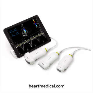
Tier 1: Handheld Ultrasound
- Description: Pocket-sized units connected to tablets or smartphones.
- Strengths:
- Quick airway marking and intubation confirmation.
- Basic gastric emptying checks in perioperative or emergency settings.
- Portable, affordable, and fast to deploy.
- Limitations:
- Lower resolution and fewer probe options.
- Limited depth and Doppler capabilities.
- Best Fit: Pre-op airway scans in OR holding areas, bedside aspiration checks in ED/ICU.
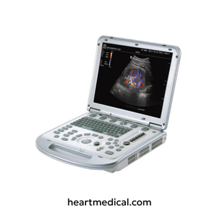
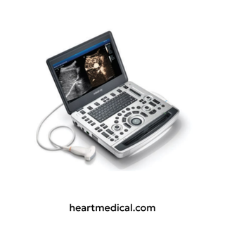
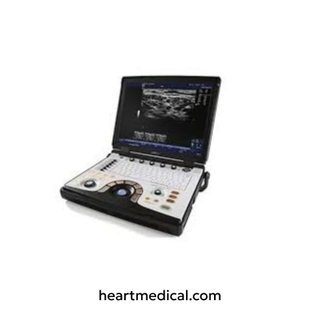
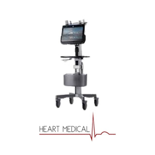
Tier 2: Portable / Compact Consoles
- Description: Laptop-style or rollable compact systems (e.g., GE Venue Go, LOGIQ e).
- Strengths:
- Higher image clarity for gastric ultrasound volume estimation.
- Multiple probe support (linear + curved + phased).
- Ideal for neuraxial pre-scans and regional blocks in addition to gastric/airway.
- Limitations: Less portable than handhelds; require stable setup.
- Best Fit: Operating rooms and anesthesia teams needing reliable, mid-range versatility.
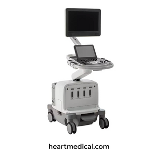
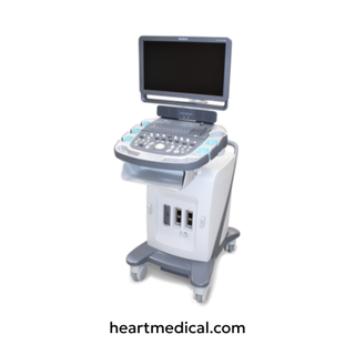
Tier 3: Advanced Cart-Based Systems
- Description: Full-sized consoles (e.g., GE Venue or premium LOGIQ).
- Strengths:
- Top-tier imaging resolution and Doppler.
- Advanced features like AI-driven needle recognition, workflow automation, and integrated archiving.
- Perfect for teaching hospitals and high-volume centers.
- Limitations: High cost, requires dedicated space and infrastructure.
- Best Fit: Academic centers, tertiary hospitals, advanced anesthesia departments.
Protocol-to-Device Fit Table
| Protocol | Handheld | Portable Console | Advanced Cart |
|---|---|---|---|
| Gastric Ultrasound | ✔ Basic empty/full check | ✔ Accurate volume estimation | ✔ High-resolution, teaching & complex cases |
| Airway Ultrasound | ✔ Cricothyroid ID & tube confirmation | ✔ Routine use with multiple probes | ✔ Advanced airway evaluation & integration |
| Neuraxial Pre-Scan | ✖ Not suitable | ✔ Good for epidural/spinal landmarks | ✔ Superior imaging, obese/difficult anatomy |
| Regional Anesthesia (UGRA) | ✖ Limited | ✔ Standard nerve blocks | ✔ Full-featured with needle recognition |
Micro-Playbooks: Step-by-Step Protocols
Gastric Ultrasound Playbook
Goal: Assess aspiration risk by identifying gastric contents.
Equipment: Curved array probe (2–5 MHz).
Steps:
- Place patient in supine, then right lateral decubitus (RLD) for best view.
- Position probe in epigastric region; identify liver (landmark) and gastric antrum.
- Observe content appearance:
- Empty: antrum flat/collapsed.
- Clear fluid: antrum round, “starry night” pattern.
- Solid: hyperechoic/heterogeneous content filling the antrum.
- Quantify if clear fluid: estimate gastric volume (low risk if <1.5 mL/kg).
- Document findings in anesthesia record.
Clinical Use: Helps guide decision on rapid sequence induction vs delay.
Airway Ultrasound Playbook
Goal: Secure safe intubation and confirm tube placement.
Equipment: Linear probe (7–15 MHz).
Steps:
- Identify Cricothyroid Membrane: Place probe transversely on anterior neck → locate hypoechoic cricothyroid membrane between thyroid and cricoid cartilage. Mark on skin.
- Predict Difficult Airway: Measure anterior neck soft tissue at hyoid/thyrohyoid level; thicker tissue suggests difficult laryngoscopy.
- Confirm Intubation: Place probe longitudinally over trachea:
- One bright-air column = tracheal intubation.
- Double air column = esophageal misplacement.
- Optional: Assess vocal cord mobility during extubation planning.
- Record confirmation for documentation.
Clinical Use: Improves safety in high-risk intubations; faster confirmation than auscultation or capnography in emergencies.
Training, QA, and Governance
Training & Competency
- Structured Learning: Start with supervised scans under experienced faculty, using standardized checklists.
- Image Repetition: Minimum recommended scans per protocol (e.g., 20–30 gastric scans, 10+ airway confirmations) before independent use.
- Device Familiarity: Training must include probe handling, presets (airway/gastric), and basic troubleshooting.
Quality Assurance
- Archiving: Save representative images for periodic review.
- Peer Review: Regular case discussions improve consistency and reduce variability.
- Benchmarks: Track scan times, success rates of gastric classification, and airway confirmation accuracy.
Governance & Safety
- Standard Operating Procedures (SOPs): Define when gastric and airway scans are mandatory (e.g., uncertain NPO status, obese patients, predicted difficult airway).
- Escalation Protocols: Ensure decisions from ultrasound (e.g., delay induction, rapid sequence intubation) are integrated into perioperative workflows.
- Compliance: Align practice with anesthesia society recommendations and infection-control guidelines (probe covers, sterile gel).
From assessing aspiration risk with gastric ultrasound to securing the airway with airway ultrasound, perioperative imaging has become central to anesthesia safety. These protocols reduce uncertainty, prevent complications, and build confidence in decision-making.
The right device tier amplifies this value, handhelds for rapid airway checks, portable consoles for everyday anesthesia workflows, and advanced cart-based systems for teaching centers and high-volume blocks. Combined with structured training and governance, anesthesia ultrasound is no longer optional; it is a standard of care.
Ultrasound Machines for Anesthesia
At Heart Medical, we offer a full collection of ultrasound machines for anesthesia backed by warranty and supported with original replacement parts, all probe types, and worldwide shipping. Whether you’re looking for new, demo, or certified refurbished systems, we have solutions tailored to your needs. We also provide the flexibility to rent ultrasound machines, making it easier for clinics and hospitals to access the technology they need without heavy upfront costs.
Reviewed by Heart Medical Clinical Applications Team
Clinical and technical specialists ensuring accuracy and relevance across all Heart Medical content.