Description
Get High Quality Imaging For Advanced Diagnosis
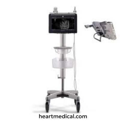
The GE Venue Fit is an advanced, compact ultrasound system tailored for critical and emergency imaging. Designed to simplify diagnostics and streamline treatment, the Venue Fit combines portability, high-resolution imaging, and AI-powered automation, making it ideal for clinicians.
Despite its lightweight design, the Venue Fit packs robust imaging features. Powered by SRI-HD speckle reduction, it delivers sharp, high-contrast images of cardiac, lung, abdominal, musculoskeletal, and vascular assessments. Its 14-inch high-definition multi-touch screen minimizes clutter for focused image analysis, while the customizable on-screen keyboard and streamlined home screen improve efficiency in high-pressure settings.
Automation is at the heart of the Venue Fit ultrasound system. Tools like Auto Optimization, Real-Time Auto Doppler, and optional Lung Sweep and Lung Scoring simplify complex tasks, reduce operator variability, and enhance reproducibility. Visual documentation tools like eFAST, renal and lung diagrams, and a patient follow-up tool support longitudinal care and clinical decision-making.
With integrated wireless connectivity, USB and Ethernet ports, and full DICOM® compatibility, the Venue Fit ensures seamless data sharing and secure archiving. Its built-in battery, power status indicators, and lightweight design make it easy to transport between patient rooms. Make your emergency cardiac assessments precise to figure out accurately what care is needed.
Key Features
1. Imaging & Performance
-
cSound™ Imageformer Technology – Delivers high-quality, poppable images with adaptive beamforming for accurate diagnostics.
-
CrossXBeam™ Compounding – Combines multiple frames from different angles to enhance contrast resolution and tissue differentiation.
-
SRI-HD (Speckle Reduction Imaging – High Definition) – Enhances image clarity by reducing speckle noise.
-
Raw Data Processing – Enables post-acquisition image optimization without degradation.
-
Virtual Convex & Virtual Apex Imaging – Expands field of view on linear and phased array probes.
-
VenueView Panoramic View – Allows extended field-of-view imaging for better anatomical context.
-
Lung Sweep Mode (optional) – Offers real-time panoramic lung imaging with cine capture for easy assessment.
-
Needle Recognition Technology – Improves needle visualization during interventional procedures.
-
Real-Time Auto Doppler Calculations – Streamlines workflow with automatic spectral Doppler measurements.
-
Simultaneous Imaging Modes – Supports duplex and triplex modes depending on probe (e.g., B/CFM/PW, B/M, Dual B + PDI).
2. User Interface & Ergonomics
-
Full Touch Interface – Intuitive multi-touch UI with tap, pinch, and swipe gestures for fast operation.
-
14" High-Resolution LCD Display – 1920 x 1080 resolution with 16:9 ratio, +/-80° viewing angle, and sealed design for durability.
-
Simple Screen Mode – Expands image display by minimizing on-screen controls; toggle UI elements with a tap.
-
Alphanumeric On-Screen Keyboard – Customizable, supports 14 languages, enables efficient data entry.
-
Home Screen Workflow – Streamlined access to clinical tools and application presets.
-
Rear Support Stand – Adjustable for flat-surface use (optional).
3. Advanced Automation & AI Tools
-
Auto Optimization – Automatically adjusts image settings for optimal clarity.
-
Lung Scoring (optional) – Enables quantitative lung evaluation.
-
eFAST, Renal, and Lung Diagrams – Visual documentation tools for point-of-care assessment.
-
Probe Assessment Tool (PAT) – Performs routine checks on probe functionality and element integrity.
-
Patient Follow-Up Tool – Enables tracking and comparison of prior scans.
-
Editable Calculations & Presets – User-defined settings and measurements for repeatability.
4. Connectivity & Data Management
-
USB 3.0 & Ethernet Ports – Three rear USBs, one side USB, and RJ45 Ethernet for fast data transfer.
-
Wireless LAN (Wi-Fi) – Enables remote access and data sharing with signal strength indication.
-
DICOM® 3.0 Compatibility – Supports Store, Print, Query/Retrieve MPPS, and Structured Reporting.
-
RSVP Remote Service – Real-time support and system updates through GE’s remote access system.
-
Integrated Archive – >60 GB storage with cine loop playback and patient data encryption.
-
Electronic Documentation Access – eIFU is available online; onboard user manual included.
5. Design & Portability
-
Compact & Lightweight Design – Weighs 5.5 kg (12 lbs) including battery and handle.
-
Optional Cart – Height-adjustable with VESA mounting, probe holders, and accessory compartments.
-
Battery & AC Power Indicators – Displayed even when the system is off; battery time remaining shown.
-
Sealed Display & Cleaning Mode – Water-resistant screen with screen cleaning capability.
Technical Specifications
1. System Architecture & Processing
-
Operating System – Microsoft® Windows® 10
-
Image Processing – Raw data processing
-
Beamforming Technology – cSound™ adaptive beamformer
-
Storage – 128 GB SSD (archive size >60 GB standard)
-
Processor – Integrated architecture optimized for point-of-care imaging
2. Dimensions & Weight
Main Unit Dimensions:
- Height: 319 mm (12.6 in.) including handle
- Width: 399 mm (15.7 in.)
- Depth: 70 mm (2.7 in.), 93 mm (3.7 in.) with handle
- Weight: 5.5 kg (12 lbs)
3. Power & Electrical Specifications
-
Power Input – AC adapter
-
Battery – Integrated battery (runtime dependent on use)
-
Indicators – Power source and battery indicators are visible when the system is powered off
4. Display & Imaging Interface
-
Main Display – 14” Multi-touch LCD
-
Resolution – 1920 x 1080 pixels
-
Viewing Angle – ±80° horizontal/vertical
-
Frame Rate – 60 Hz
-
Audio – Integrated speaker with volume control
5. User Interface & Control
-
Touchscreen – Multi-touch with gesture recognition
-
Customizable Presets – User-defined exam and control settings
-
Thumbnails & Viewer – Integrated image viewer for quick review
-
Annotations & Body Marks – Editable, application-specific tools
-
Onboard Keyboard – Alphanumeric touch keyboard
6. Connectivity & Storage
-
USB Ports – 3 rear, 1 side (USB 3.0)
-
HDMI Output – Compatible with external displays
-
Ethernet – RJ45 (10/100/1000 Base-T)
-
Wireless LAN – Wi-Fi enabled with signal strength display
-
Data Export – DICOM, JPEG, MPEG, and anonymized formats
-
Patient Data Security – Encryption and anonymization supported
Probe Compatibility
1. Phased Array Probes (Cardiac & General Imaging)
These compact probes offer deep penetration and real-time imaging, ideal for cardiac, abdominal, and pediatric assessments in point-of-care and emergency settings.
-
3Sc-RS (1.3 – 4.0 MHz) – Suitable for cardiac, pediatric, abdominal, and adult cephalic imaging. Also compatible with LVO contrast imaging (optional).
-
6S-RS (2.0 – 7.0 MHz) – Optimized for pediatric, fetal, neonatal cephalic, and abdominal applications.
-
12S-RS (4.5 – 12.0 MHz) – High-resolution probe ideal for pediatric and neonatal cephalic imaging.
2. Transesophageal (TEE) Probe (Cardiac Imaging)
TEE probes enable detailed cardiac visualization via the esophagus, especially in critical care and intraoperative scenarios.
3. Convex & Micro Convex Array Probes (Abdominal, OB/GYN & Urology Imaging)
These probes provide wide field-of-view and deep penetration for abdominal, fetal, and pelvic assessments.
-
C1-5-RS (1.5 – 5.0 MHz) – Used for general abdominal, OB/GYN, fetal, and urological imaging.
-
8C-RS (3.5 – 10.0 MHz) – Microconvex probe ideal for neonatal, pediatric, and abdominal scanning with superior detail resolution.
-
E8C-RS (3.5 – 11.0 MHz) – Versatile micro convex probe for OB, GYN, fetal echo, prostate, and transrectal imaging.
4. Linear Array Probes (Vascular, MSK & Small Parts Imaging)
These high-frequency probes are suited for superficial imaging including vascular access, MSK evaluations, and small organ imaging.
-
9L-RS (3.0 – 10.0 MHz) – Supports peripheral vascular, abdominal, pediatric, and musculoskeletal imaging.
-
12L-RS (4.0 – 13.0 MHz) – Used for vascular, small parts, and neonatal cephalic imaging.
-
L4-12t-RS (4.0 – 12.0 MHz) – Compact linear probe designed for MSK, vascular, and procedural guidance.
-
L8-18i-RS (4.5 – 18.0 MHz) – High-frequency imaging for small parts, superficial MSK, and intraoperative applications.
-
L10-22-RS (5.0 – 22.0 MHz) – Ultra high-frequency probe for dermatology, superficial MSK, and small structure imaging.
-
L4-20t-RS (XDclear) (4.0 – 20.0 MHz) – Advanced XDclear probe with improved image quality and penetration, designed for vascular, MSK, and interventional procedures.
5. Linear Array with Active Matrix (Small Parts & High-Resolution Imaging)
Matrix technology enhances beam focusing, resulting in superior resolution and uniformity.
-
ML6-15-RS (4.0 – 15.0 MHz) – Active Matrix probe for breast, thyroid, small parts, and MSK imaging, delivering excellent near-field resolution.
Applications
1. Cardiac Imaging (Adult & Pediatric)
Essential for emergency, critical care, and point-of-care cardiac assessments.
- 2D Cardiac Imaging for adult and pediatric hearts.
- Focused Cardiac Ultrasound (FoCUS) for rapid bedside evaluations.
- Left Ventricular Function Assessment for detecting cardiac dysfunction.
- Transesophageal Echocardiography (TEE) for detailed heart structure visualization (with 6Tc-RS).
2. Abdominal & General Imaging
Supports rapid and accurate assessment of abdominal organs and pathologies.
- Liver, Gallbladder, Kidney, and Pancreas Imaging with high-resolution visualization.
- Aorta and Renal Artery Evaluation for detecting aneurysms, stenosis, or obstructions.
- Focused Assessment with Sonography for Trauma (FAST) in emergency settings.
3. Thoracic & Pleural Imaging
Fast evaluation of lung conditions and pleural abnormalities.
- Detection of Pleural Effusion, Pneumothorax, and Lung Consolidation.
- Assessment of Diaphragmatic Motion and Lung Sliding.
4. Neonatal & Pediatric Imaging
High-frequency probes provide clarity for small anatomical structures.
- Neonatal Head, Abdomen, and Hip Imaging for congenital and developmental evaluations.
- Pediatric Abdominal and Cardiac Assessments with optimized probes.
5. Adult & Neonatal Cephalic Imaging
Detailed cranial imaging for early diagnosis and monitoring.
- Cranial Ultrasound in neonates for intraventricular hemorrhage and brain development assessment.
- Cephalic Views in adults for soft tissue and sinus evaluations.
6. Breast & Small Parts Imaging
High-resolution imaging for screening and diagnostics.
- Breast Ultrasound for cysts, masses, and guided interventions.
- Thyroid, Testicular, and Soft Tissue Imaging for nodules, lesions, and vascularity.
7. Gynecology & Obstetric Imaging
Essential for reproductive health and prenatal care.
- Transvaginal and Transabdominal Scans for uterine and ovarian evaluation.
- Fetal Biometry, Growth Tracking, and Amniotic Fluid Assessment.
- Follicular Monitoring and Early Pregnancy Detection.
8. Musculoskeletal (MSK) Imaging
Accurate soft tissue and joint assessments for injury and pain management.
- Tendon, Ligament, and Joint Space Visualization.
- Soft Tissue Lesion Evaluation and Dynamic Movement Assessments.
9. Nerve Imaging & Guidance
- Used in anesthesia and pain management procedures.
- Real-Time Peripheral Nerve Localization.
- Guidance for Nerve Blocks and Regional Anesthesia.
10. Vascular & Vascular Access Imaging
- Supports vascular assessment and procedural guidance.
- Color and Spectral Doppler Imaging for vessel patency and flow dynamics.
- Peripheral Artery and Vein Evaluation for DVT, stenosis, and thrombosis.
- Central and Peripheral Vascular Access Guidance in real-time.
11. Urological & Prostate Imaging
- Reliable imaging for urological assessments and prostate screening.
- Bladder Volume Measurement and Retention Evaluation.
- Transrectal Prostate Imaging (TRUS) for gland size and lesion detection.
- Kidney Imaging and Doppler Assessment for perfusion studies.
Frequently Asked Questions
1. What operating system does the GE Venue Fit use?
The GE Venue Fit operates on a Windows 10 platform, offering a secure, intuitive interface with fast boot-up, seamless connectivity, and easy integration into hospital networks and electronic health records (EHR) systems.
2. Is the GE Venue Fit designed for point-of-care and emergency settings?
Yes, the Venue Fit is optimized for Point-of-Care Ultrasound (POCUS), making it ideal for critical care, emergency, and bedside applications. Its intuitive interface, quick startup, and portable design cater specifically to fast-paced clinical environments.
3. Does Venue Fit support needle visualization for guided procedures?
Yes, GE's Advanced Needle Visualization technology enhances needle visibility during interventional procedures, improving safety and accuracy in vascular access, biopsies, and regional anesthesia.
4. Does the Venue Fit have remote support capabilities?
Yes, it includes InSite™ remote support functionality (via secure GE Healthcare access), enabling real-time diagnostics, troubleshooting, software updates, and remote training.
5. Are automated tools available for faster and more accurate assessments?
Yes, Venue Fit is equipped with AI-powered tools like Auto VTI, Auto B-Lines, and Auto IVC for enhanced speed and consistency.
6. What are the data storage and image transfer options available?
- The Venue Fit provides flexible storage and export capabilities:
-
Solid-state drive (SSD) for fast internal data access and storage.
-
USB ports for exporting studies to memory sticks or external drives.
-
DICOM connectivity for PACS integration, including DICOM Store, Modality Worklist, and Structured Reporting (SR).
- Wi-Fi and Ethernet for network connectivity and data sharing.