POCUS ultrasound (Point of care) has matured into a standards-based bedside toolkit: reproducible protocols (e.g., eFAST for trauma; BLUE for acute respiratory failure; RUSH for shock; focused cardiac POCUS for rapid hemodynamic reads) let clinicians answer binary questions fast and safely at the point of care.
This article narrows to two levers that drive outcomes and governance:
- Protocol discipline: What each canonical exam answers, common pitfalls, and how scans ladder into decision trees; and
- Device tiers, portable/compact, and advanced carts: Mapped to protocol demands (image quality, Doppler, probe set, uptime), with evidence that modern handhelds can meet many protocol needs while consoles still dominate advanced quantification.
We’ll anchor to major society guidance and contemporary reviews, then give a protocol-to-device matrix you can operationalize at the bedside, QA, and training levels.
Core POCUS Ultrasound Protocols Every Clinic Should Know First
Below are two high-impact protocols that cover urgent diagnostic needs. Mastering these gives confidence and broad applicability across clinical settings.
1. eFAST / FAST (Trauma Protocol)
What It Answers:
- Is there free fluid in the abdomen (hemoperitoneum)?
- Is there air or fluid in the chest (pneumothorax, hemothorax)?
- Is there pericardial effusion (tamponade)?
Views / Steps:
- Right Upper Quadrant (RUQ): Morison’s pouch, between liver and kidney.
- Left Upper Quadrant (LUQ): Spleen, splenorenal area.
- Pelvic view: Suprapubic, pouch of Douglas / rectovesical depending on sex.
- Cardiac / Subxiphoid view: For pericardial fluid / tamponade.
- Bilateral lung / chest views: For pneumothorax / hemothorax.
Benefits & Use Cases:
- Rapid screening in trauma settings.
- Helps triage patients for CT or surgery when they are unstable.
Limitations / Pitfalls:
- Low sensitivity for small internal injuries or bowel injuries without free fluid.
- Operator-dependent: body habitus, presence of air, and skill matter.
- Negative eFAST does not rule out serious injury; clinical monitoring or repeat scans may be needed.
2. Lung / BLUE Protocol (Acute Respiratory Distress / Dyspnea)
What It Answers:
- Is there lung consolidation, pleural effusion, pulmonary edema?
- Are signs of pneumothorax present?
- Helps differentiate causes of shortness of breath (e.g., pneumonia vs heart failure vs ARDS vs COPD exacerbation).
Views / Steps:
- Anterior chest: Look for lung sliding, B-lines, A-lines.
- Lateral and posterior lung regions for consolidation or effusions.
- Assess for pleural fluid above diaphragm.
Benefits & Use Cases:
- Immediate bedside help when chest X-ray or CT is delayed or unavailable.
- Helps guide management in ICU, ER, or outpatient acute care.
Limitations / Pitfalls:
- Less effective in obese or COPD patients due to poor acoustic windows.
- Interpretation requires pattern recognition training.
Why These Protocols First
- They address life-threatening, common emergencies (trauma, respiratory failure).
- They require only a minimal set of probes (often phased-array or curvilinear + linear).
- They can be executed quickly (often under 5 minutes for eFAST) and repeatedly.
Device Tiers for POCUS: Matching Protocols to Equipment
Philips and other manufacturers all offer solutions that fall into three clear tiers: handheld, portable/compact consoles, and advanced cart-based systems. Each tier carries strengths and limitations depending on the protocol.

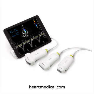
Tier 1: Handheld / Ultra-Portable
- What They Are: Small, pocket-sized devices that connect to a phone or tablet.
- Best For: Rapid bedside checks, trauma (eFAST), lung ultrasound, vascular access.
- Strengths: Portability, affordability, instant boot-up, suitable for emergency and field settings.
- Limitations: Lower image depth, limited probe selection, not ideal for detailed cardiac quantification.
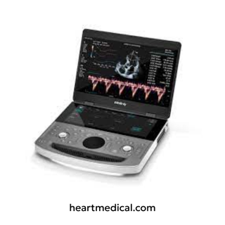
Tier 2: Portable / Compact Consoles
- What They Are: Laptop-style or compact systems with broader probe support.
- Best For: Multi-protocol use (eFAST + Lung + FoCUS), emergency departments, critical care, and clinics with moderate patient volume.
- Strengths: Higher image quality than handhelds, Doppler support, wider probe selection.
- Limitations: Less portable than handhelds; higher cost; require stable power sources.
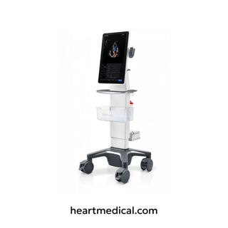
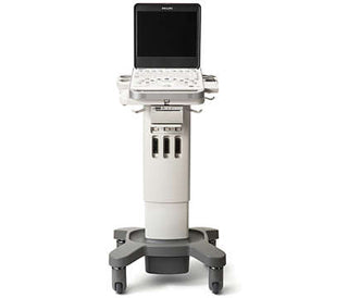
Tier 3: Advanced Cart-Based Systems
- What They Are: Full-size consoles with the most advanced features.
- Best For: High-volume hospitals, teaching centers, or specialty clinics needing advanced quantification.
- Strengths: Superior image resolution, advanced Doppler, 3D/4D capabilities, broadest protocol coverage.
- Limitations: Expensive, less portable, requires dedicated space and trained operators.
Protocol-to-Device Fit Snapshot
| Protocol | Handheld | Portable Console | Advanced Cart |
|---|---|---|---|
| eFAST / Trauma | ✔ Rapid screening | ✔ Broader probe set | ✔ High-res for complex trauma |
| Lung / BLUE | ✔ Bedside checks | ✔ Clearer lung/pleural views | ✔ Teaching & advanced imaging |
| Focused Cardiac (FoCUS) | ✖ Limited | ✔ Adequate for routine FoCUS | ✔ Ideal for full cardiac studies |
| RUSH / Shock | ✖ Partial views only | ✔ Balanced portability & Doppler | ✔ Best for advanced hemodynamic workups |
Looking for a POCUS Ultrasound?
At Heart Medical, we help clinicians choose the right POCUS solution backed by authorized access to:
- New, demo, and certified refurbished systems tailored to your protocols.
- Full probe and accessory support, so your device grows with your clinical needs.
- Warranty and service coverage that keeps downtime to a minimum.
Reviewed by Heart Medical Clinical Applications Team
Clinical and technical specialists ensuring accuracy and relevance across all Heart Medical content.